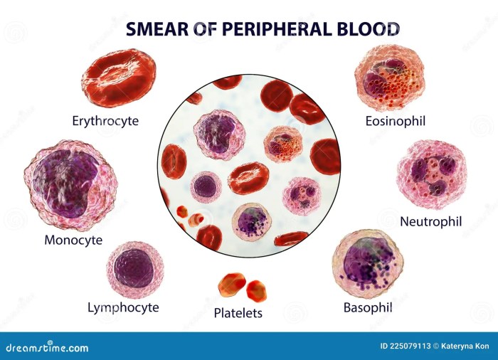
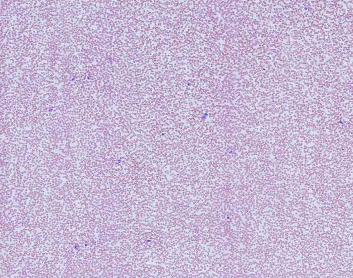
Figure 20.7 Peripheral Blood Smear presents a comprehensive overview of the cellular components found in a peripheral blood smear, providing a detailed examination of their morphology and characteristics. This in-depth analysis serves as a valuable diagnostic tool for hematological and systemic disorders, aiding in the identification and differentiation of various conditions.
This Artikel explores the normal findings, pathological variations, technical aspects of preparation, clinical applications, and differential diagnosis using peripheral blood smear analysis, offering a thorough understanding of this essential laboratory technique.
Peripheral Blood Smear Analysis: Figure 20.7 Peripheral Blood Smear

Peripheral blood smear analysis is a diagnostic tool used to evaluate the cellular components of blood. It involves spreading a thin layer of blood onto a glass slide and staining it to differentiate between different types of cells. This technique provides valuable insights into hematological disorders, systemic conditions, and overall health status.
1. Peripheral Blood Smear Components, Figure 20.7 peripheral blood smear
A peripheral blood smear consists of various cell types, each with unique morphology and characteristics:
- Red Blood Cells (RBCs):Erythrocytes are non-nucleated, biconcave discs that carry oxygen. Their size, shape, and hemoglobin content can provide information about anemia, hemolytic disorders, and other conditions.
- White Blood Cells (WBCs):Leukocytes are nucleated cells responsible for immune defense. Different types of WBCs include neutrophils, lymphocytes, monocytes, eosinophils, and basophils, each with specific roles in the immune system.
- Platelets:Thrombocytes are small, non-nucleated fragments of cells that play a crucial role in blood clotting.
2. Normal Findings in a Peripheral Blood Smear
The normal ranges for different cell types in a healthy individual are:
| Cell Type | Normal Range |
|---|---|
| Red Blood Cells | 4.5-5.9 million/μL |
| White Blood Cells | 4,000-11,000/μL |
| Neutrophils | 50-70% |
| Lymphocytes | 20-40% |
| Monocytes | 3-8% |
| Eosinophils | 1-3% |
| Basophils | <1% |
| Platelets | 150,000-450,000/μL |
Deviations from these ranges can indicate underlying conditions such as infections, anemia, or leukemia.
3. Pathological Findings in a Peripheral Blood Smear
Abnormal cell morphologies and variations in a peripheral blood smear can help diagnose specific diseases or conditions:
| Pathological Finding | Associated Condition |
|---|---|
| Anemia (low RBC count) | Iron deficiency, blood loss, chronic diseases |
| Leukocytosis (high WBC count) | Infections, inflammation, leukemia |
| Neutrophilia (high neutrophil count) | Bacterial infections, acute inflammation |
| Lymphocytosis (high lymphocyte count) | Viral infections, chronic infections, leukemia |
| Monocytosis (high monocyte count) | Chronic infections, autoimmune disorders |
| Eosinophilia (high eosinophil count) | Parasitic infections, allergic reactions |
| Basophilia (high basophil count) | Chronic myelogenous leukemia, allergic reactions |
Commonly Asked Questions
What is the significance of normal findings in a peripheral blood smear?
Normal findings in a peripheral blood smear establish a baseline for healthy individuals, providing reference values for each cell type. Deviations from these ranges can indicate underlying conditions, prompting further investigation and appropriate medical intervention.
How does peripheral blood smear analysis contribute to differential diagnosis?
Peripheral blood smear analysis plays a crucial role in differential diagnosis by examining specific cell morphology changes associated with various hematological conditions. By comparing the observed abnormalities to known patterns, healthcare professionals can narrow down the diagnostic possibilities and guide further testing.
What are the key technical aspects to consider when preparing a peripheral blood smear?
Proper sample collection and handling are essential for obtaining a high-quality peripheral blood smear. This includes using appropriate anticoagulants, maintaining the correct blood-to-anticoagulant ratio, and creating a thin, even film on the glass slide. Following standardized techniques ensures accurate and reliable results.

